

The area near his hip where the lump was located, has less spare skin, which can make the surgery more challenging. Greyhounds are also naturally thin skinned and this can make them more prone to problematic wound closure.
Because the surgery was to be extensive, Louise referred Ollie to a soft tissue specialist.
Ollie's operation went well; the first stage was to remove the tumour, before the surgeon used a skin flap technique to close the large remaining wound.This was the best option for bringing the wound edges together under the least amount of tension. Post operatively, Ollie spent a few days in hospital at the referral centre. He was a good boy, apart from when he managed to take his buster collar off and nibble at a stitch! The nibbled stitch was at the tip of the flap that had the worst blood supply, so there was already a concern that this part of the wound might not hold together.

Ollie was allowed home on strict rest, but on day three of being home, Ollie decided to have a roll around on the floor and the wound opened up further (wound picture1). Louise spoke to the specialist and it was decided that the best course of action was to attempt to re-close the wound. Louise carried out the procedure.With Ollie anaesthetised, she flushed the wound and closed it using special tension relieving stitches called walking sutures.For Ollie's wound to have the best chance of healing he needed to be kept quiet and rested, so he was to spend the next 10 - 14 days in hospital.Ollie was given pain relief medication to help keep him comfortable as well as antibiotics. He also had to wear a buster collar again so that he couldn't lick the area. The nurse's gave him lots of cuddles as he was understandably feeling rather sorry for himself.Ollie's wound was monitored closely; it was beginning to heal, apart from the one problem area at the top of the wound. Senior nurse Dayna recommended daily dressings using medical grade manuka honey that supports the healing process (wound picture 2). He was such a brave boy as it must have been sore having it dressed.

The area on Ollie's thigh was difficult to bandage, so to help keep the dressing in place, he wore a special body bandage. Ollie was doing well and his wound was looking better, when he unfortunately suffered a set back.
Ollie managed to remove the buster collar and body bandage that he'd been comfortably wearing, licked the wound and opened it up again!
Ollie was referred back to the specialist centre and he was admitted to the hospital for open wound management.They attached his buster collar to a special harness so that he couldn't get it off.
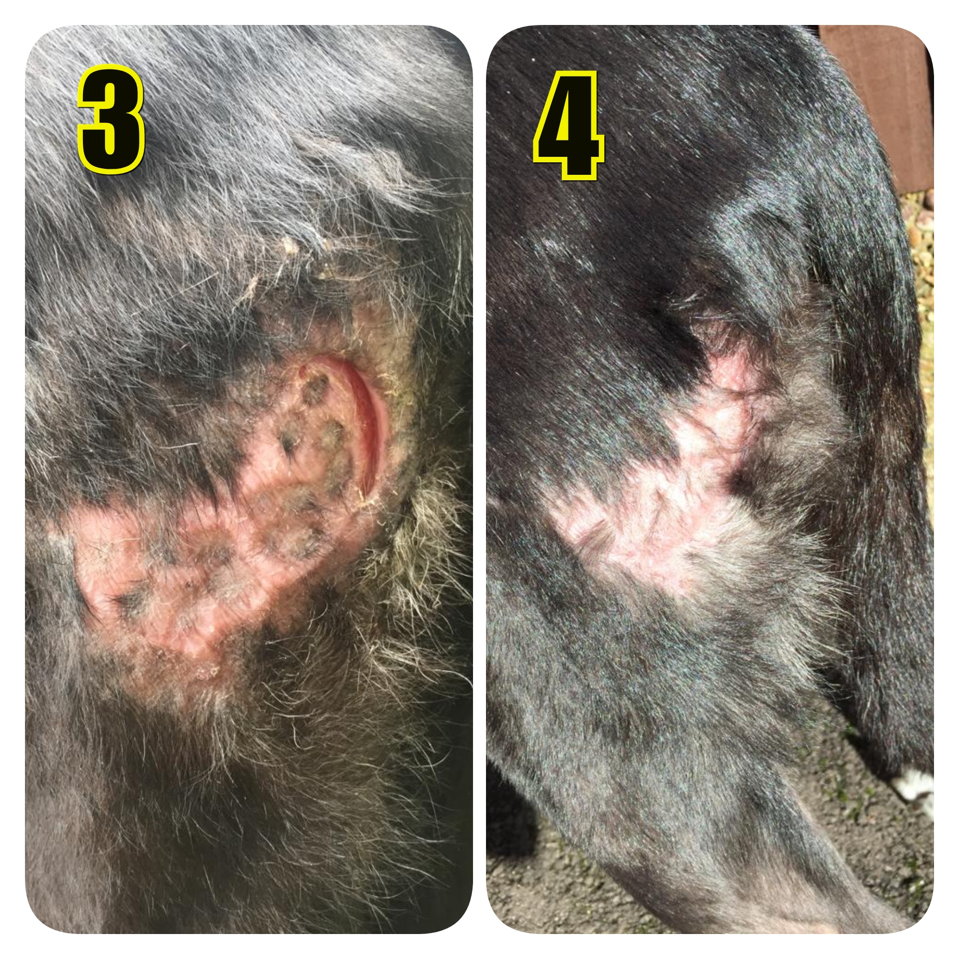 Although his wound was looking healthy, it was taking a long time to heal, so the decision was made to perform punch skin grafts. To do this, Ollie was given a general anaesthetic. An area on his abdomen was clipped and cleaned to allow the surgeon to remove small pieces of circular skin from the site. He then placed them into the granulation tissue ( healing tissue) of the wound. It was hoped that the majority of the punched biopsies would 'take' allowing new skin to build between them (epithelialisation).Ollie recovered well from the procedure and continued with strict cage rest. Three weeks later Ollie was allowed home from hospital. Louise continued to dress the wound every three days. Healthy pink epithelial tissue formed ( wound picture 3) and finally his wound healed (wound picture 4).There is no fur on the skin over the grafted areas as it came from the hairless skin on his tummy!
Following analysis of the tumour at the laboratory, the results were encouraging. The surgeon had managed to remove the whole tumour and immunohistochemistry testing gave it a low grade result.There is a low chance of recurrence so to be on the safe side we will monitor Ollie regularly.
It's been a long road to recovery and we are so pleased to see that Ollie is his happy self again and enjoying his most favourite thing - walks with his friend, Nigel the Whippet!
For his patience and bravery, Ollie is our much deserved pet of the month!
