
Tuesday, 21 August 2018

Pet of the month Hall of fame - Loki (June 2018)

During the spring bank holiday (28th May), Loki had fallen from a height of about 50cm and hit his head. He appeared confused and was reluctant to move, so his owner called our emergency service and vet Karolina who was on duty arranged to see him straight way. Loki was given a thorough examination and fortunately he had not injured his back and neck, and he was alert and able to move around.  However, he had a small puncture wound on his lip and a sore jaw that he wouldn’t allow Karolina to touch. Karolina gave Loki an injection of pain relief and allowed him home for the night so that he could rest in his own familiar surroundings. She arranged to see him again the following morning. Loki was happier but his jaw was still sore and he was having trouble eating his nugget food. Karolina could now see that his lower jaw was broken in between his two front teeth, an area called the mandibular symphysis, where the bones of the right and left mandible meet. This meant that his jaw was unstable and preventing him from chewing his food –something that rabbit’s like to do all day. It is also vital for their health; Rabbits thoroughly chew and grind their food so that their teeth, that are continually growing, are worn down and renewed. Loki required a general anaesthetic so that his jaw could be realigned and the fracture stabilised.

Vet Kirstie anaesthetised Loki and firstly took an x-ray of his skull to check for other injuries and to assess the lower jaw injury. The next stage was to meticulously realign the lower jaw bones before using a special tissue glue to glue the two lower incisors together. This effectively stabilised the jaw. She then used a thick suture material to further secure the jaw fracture in place. 


Loki recovered well from the general anaesthetic and it was important that we provided nutritional support throughout his recovery until he was able to eat well enough by himself. At home Loki has a hutch mate called Poppy whom he lives with, so to ensure neither of them became lonely, Poppy came to stay. Loki was a much happier bunny and managed to eat some grass and greens by himself. Loki was allowed home the following day and he continued to do really well.


Six weeks after his operation, Loki came to see Kirstie and she was so impressed with his progress that it was time to remove the suture material. Loki was given a short acting anaesthetic to enable Kirstie to do this. The glue that had been used just disperses naturally as the teeth grow.


We are all so pleased to see that this gorgeous boy is better. For being a brave and tolerant patient and for his cuteness, Loki is a much deserved pet of the month! 


Malocclusion is a complication that can develop following an injury like Loki's. This is where poor teeth alignment lessens the effectiveness of chewing and leads to overgrown teeth. It can inhibit their ability to eat. Rabbit's teeth can also become overgrown because of a poor diet or due to malocclusion present from birth.

Loki's owner will continue to monitor him closely to ensure that he is eating.


Friday, 20 July 2018

Pet of the Month Hall of Fame - Jeffery ( May 2018)

The Great Dane is an unmistakeable breed. They may be a large dog but they are often referred to as gentle giants and six year old Jeffery is no exception.
Like other large breed dogs, they are at risk of a life threatening form of bloat. 
Gastric dilatation - volvulus (GDV) is a veterinary emergency which occurs as a result of the stomach filling up with gas (a bit like a balloon), becoming increasingly distended, and the gas cannot escape (dilatation). The stomach can also twist on itself impairing blood flow (volvulus).
This is a rapidly progressive emergency as the blood vessels on the stomach's surface become stretched, reducing their ability to supply blood to the stomach wall. As the stomach twists, it cuts off the blood flow to vital blood vessels, which can lead to the stomach tissue dying. Circulation is also impaired. The animal quickly deteriorates, becoming increasingly weak and shocked. This fatal process can happen within a few hours so it is important to spot the symptoms and seek immediate veterinary attention - surgery is the only cure. 

A few weeks ago, Jeffery's owners found themselves in this very situation. It was Sunday teatime and after finishing his dinner, Jeffery went out into the garden for a wander. His owner Karen noticed that he'd been outside for a while so went to check on him. Jeffery was pacing around the garden and he looked like he wanted to be sick. He attempted to vomit, but was unable to bring anything up apart from some thick saliva secretions
(unproductive vomiting). Jeffery was restless but he had become lethargic and kept lying down - all tell-tale signs of  a GDV. Karen managed to take Jeffery indoors where he began to cry in pain and it was then that Karen noticed that Jeffery's abdomen (belly) had become swollen and distended - a classic symptom of bloat. Recognising the situation, Karen called the hospital and got in touch with the head vet Cathy who was on duty.


From the symptoms described by Jeffrey's owner, Cathy suspected that Jeffery was suffering from GDV and arranged to meet them at the hospital straight away.

Upon arrival, Cathy examined Jeffrey and confirmed the diagnosis .With the assistance of student nurse Becky, Jeffery was given pain relief and fluid via an intravenous drip.  His owners consented to the surgery and Jeffery was admitted to the hospital. His stomach was hugely distended, so to relieve the pressure, Cathy inserted a large gauge needle through his body wall and into his stomach to allow some gas to escape. Becky set up in theatre and prepared the anaesthetic equipment . Jeffery was now stabilised  enough for him to undergo the general anaesthetic required.
Once in theatre, to surgically correct the GDV,  Cathy firstly needed to very carefully 'de-rotate' the stomach back into it's normal position. Jeffery's spleen had also twisted due to the movement of his stomach, so Cathy needed to place this back into it's normal position in the abdomen. Next Cathy assessed the tissues of his stomach and spleen and luckily the blood supply and tissues looked active and healthy. The next step was to pass a stomach tube down Jeffery's oesophagus and into the stomach in order to empty it's contents and further relieve the pressure. Unfortunately this procedure was unsuccessful due to the stomach content being too thick, so Cathy was then required to carry out a procedure called a gastrotomy in which an incision is made into the stomach wall. This way, Cathy was able to remove the stomach contents. The last part of the operation was to carry out a gastropexy. This is where the stomach is attached to the inside of the body wall, so it can no longer shift and twist. Throughout the emergency operation, student nurse Becky closely  monitored Jeffery's general anaesthetic. 

Jeffery recovered very well from the general anaesthetic, and the surgery proved to be a success.

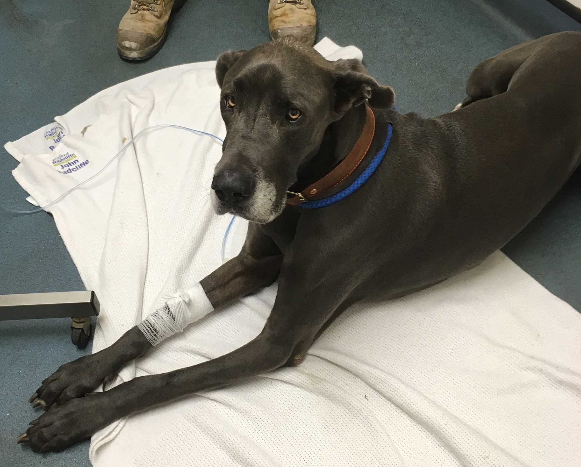
After a night in the hospital, Jeffery was looking so much happier and he had re-discovered his appetite. The nurses monitored him closely for signs of vomiting and discomfort as well as ensuring his surgical wound was clean and dry. He enjoyed having some gentle strolls around the garden and getting lots of attention from the nurses.


(Pictured with our head nurse Jazmin) .

Cathy was so pleased with Jeffery's progress that she allowed him to go home just two days after his life saving operation. Fortunately the quick thinking action of his owners meant that Jeffery was seen within an hour and an half from when his first symptoms developed - this contributed to a successful outcome. 


We are all so pleased to see that Jeffery has recovered so well and is a much happier boy!

For being so brave and for his gorgeous nature, Jeffery is a much deserved pet of the month!


The causes of GDV are not clearly known but there are several factors that appear to increase an individual's risk of developing the condition. Whilst any breed of dog and even cats can develop GDV, it is most commonly seen in large/giant breeds of dog which are deep chested. Nervous, anxious dogs under stress may be more prone to developing it, as are animals that eat a large meal  or drink a large amount of water in one go. Eating rapidly may also contribute to the animal gulping in air. Exercising just before or after a meal is another possible factor. 

In order to minimize the risk of developing GDV it is suggested that dogs should be fed frequent smaller portions of food rather than one large meal. Don't allow your dog to heavily drink in one go, especially after a meal - instead offer smaller amounts often. Strenuous exercise and excitement such as play should be avoided before and after eating.

Most importantly, be aware of the signs of a GDV - panting, discomfort, restless, pacing, lethary, drooling, unproductive attempts to vomit ( retching and only bringing up white froth ), crying and a distended abdomen ( belly ) -  bloated, full appearance.
Remember, pets don't read the text books - symptoms can vary and not all symptoms will be seen. If in any doubt, do seek immediate veterinary advice.


Disclaimer:
      The contents of the Arden House Animal Hospital website are for informational purposes only. The content is not intended to be a substitute for professional veterinary advice, diagnosis, or treatment. Always seek the advice of your Veterinary Surgeon with any questions you may have regarding your animal’s medical condition. Never disregard professional medical advice or delay in seeking it because of something you have read on this website. 


Thursday, 14 June 2018

Pet of the month - Hall of Fame, Clovie (March 2018)


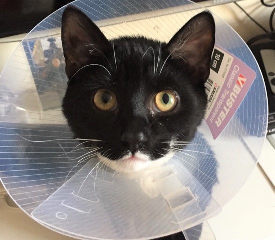
Clovie was brought in by his owners after they spotted that he’d been sick and had brought up a small piece of plastic and a small amount of thread. Vet Karolina examined Clovie and found his vital signs to be normal. He was a little quiet but bright, so initially Karolina gave him some medication to settle his stomach and allowed him to go home so that  Clovie’s owners could watched him closely for any further symptoms. Clovie was sick again so the next step was to admit him to the hospital for further tests.

Karolina placed him onto a drip and took an x-ray of his abdomen. The x-ray did not detect an obvious foreign body, however some foreign bodies are not visible on x-ray and the vet looks for changes in the gas pattern within the digestive tract. Clovie was constipated so Karolina gave him an enema which helped him to pass faeces. To Karolina's surprise, his faeces was a dark blue colour! Clovie's owners thought that the plastic might have been from a pen, and with the evidence of blue faeces, this was credible. Karolina felt his abdomen again and although it was not painful, there was one suspicious area, possibly as a result of swollen intestinal lymph nodes due to inflammation. Although Clovie was bright and he’d not been sick again,Karolina could not rule out the possibility that he’d eaten  something else that he shouldn’t have. Clovie remained in hospital on supportive treatment and the following morning vet Louise continued his care and assessed him.

Despite Clovie remaining bright, Louise was also concerned that his abdomen felt slightly abnormal. There was a risk that Clovie had eaten more thread and after a discussion with Clovie’s owners it was agreed that Clovie should have an operation called an exploratory laparotomy to allow Louise to look for a foreign body. Clovie was placed under a general anaesthetic and the nurses prepared him for theatre. Once in theatre Louise could feel something in Clovie’s stomach and she could also see that part of his small intestine was bunched up. This is a sign consistent with a linear foreign body. This term describes long, thin objects such as string, wool, thread and tinsel.

Firstly Louise carried out a procedure called a gastrotomy which involved making an incision into Clovie’s stomach. This revealed a wad of plastic and some thread. Louise removed the plastic and then assessed the thread, she could see that the thread was extending out of the stomach and into the small intestine. One end of a linear foreign body will tend to lodge itself somewhere in the gastro - intestinal tract, commonly around the base of the tongue or in the bottom of the stomach as in Clovie’s case. Even though part of the thread was stuck in his stomach, the normal movement of the small intestine had continued to move the rest of the thread onwards through them. The trouble here is, the intestines keep moving but the thread does not. This causes the intestine to plicate or ‘bunch up’ on itself. This is a particularly dangerous situation as the blood supply to the area can deteriorate causing the tissue to die. In some cases the linear foreign body can tear through the tissue of the intestine, leaking its contents into the abdominal cavity, resulting in a severe infection called peritonitis.

Fortunately for Clovie it had not reached this stage and although his small intestine was inflamed, it looked healthy. The next stage of the operation was for Louise to remove the thread. In order to safely do this, Louise needed to make three separate incisions along the small intestine (enterotomies). She then carefully followed the thread along, cutting it before gently removing it through each incision. Upon completion of the operation Clovie recovered well from the anaesthetic and the next day was feeling much better. He was asking for cuddles, of course we happily obliged! To encourage him to eat, the nurses’ hand fed him and he was soon tucking into the food by himself. Clovie has recovered very well and we are so pleased to see him back home and of course, the centre of attention.

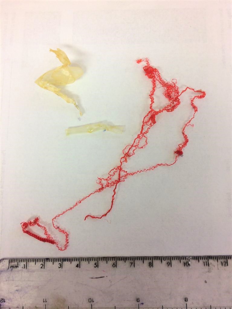
The plastic and thread that Clovie had eaten!


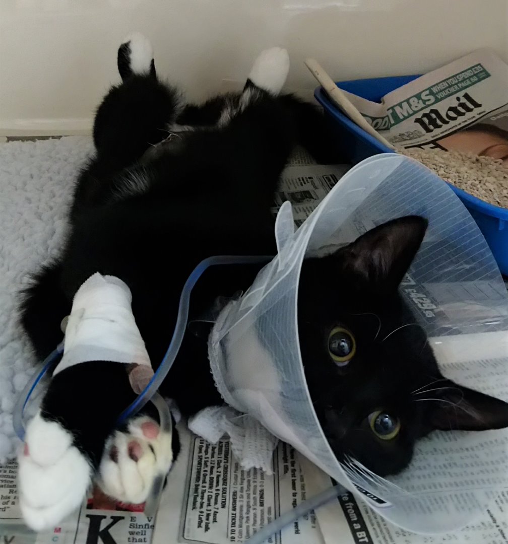
Clovie the day after his life saving operation.


Clovie's owners discovered that the thread had come from his favourite thing to play with, a cat Santa hat. Part of the thread had become exposed and unfortunately unravelled.  Cats don’t often intentionally mean to swallow thread, it gets caught on the rough barbs called papillae on their tongue and they can’t spit it out. They may try to use a paw to get it out but if this isn’t successful they end up swallowing it.

The plastic was identified as the inner coating of a felt tip pen!

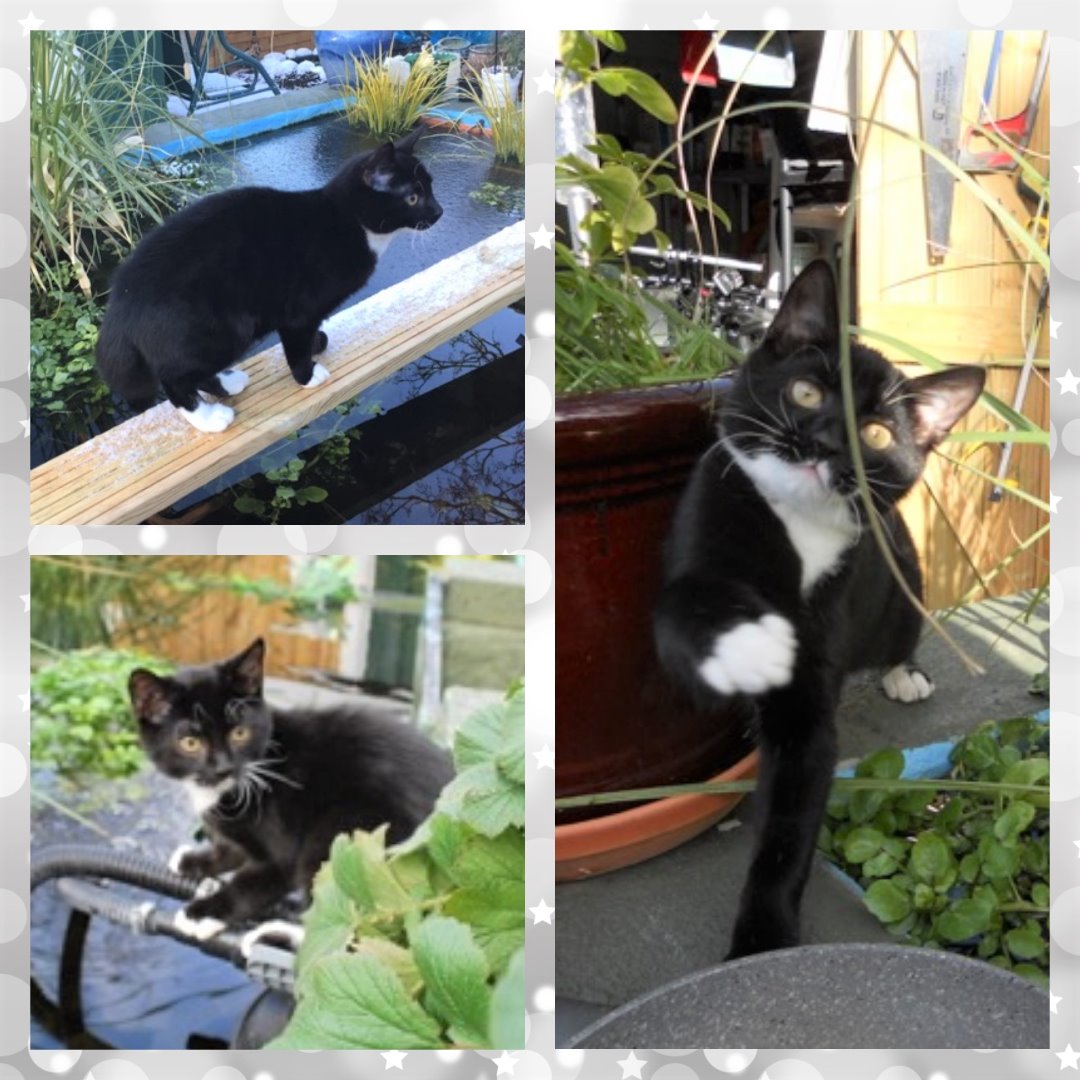
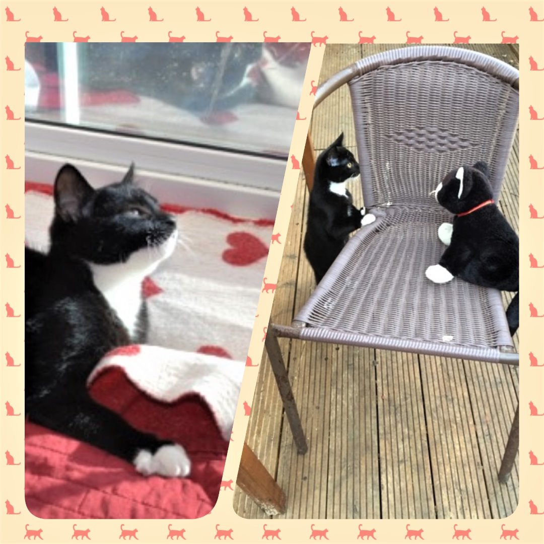
Thank you to Clovie's owners for allowing us to share his story. They sent us in these super photos with this lovely message;


'Thank you for picking our Clovie to be Pet of the month. As you know he was a poor little boy but thanks to all of you we have got our baby back to his lively happy self, and we are so very grateful. '


 Many thanks,

Elena and Glyn.


  Disclaimer  
The contents of the Arden House Animal Hospital website are for informational purposes only. The content is not intended to be a substitute for professional veterinary advice, diagnosis, or treatment. Always seek the advice of your Veterinary Surgeon with any questions you may have regarding your animal’s medical condition. Never disregard professional medical advice or delay in seeking it because of something you have read on this website